A Maximum Intensity Projection (MIP) is a series of 2D images generated from a 3D reconstruction by selecting the maximum value through the reconstruction seen by each 2D pixel at each of several viewing angles.
Each angular view in a MIP has a unique viewing angle which defines the 2D projection plane. Parallel rays emanate from each of the pixels in the projection plane through the reconstruction. The maximum intensity, i.e. largest-valued voxel, encountered by each ray is assigned to the pixel from which that ray emanated. For example, bone typically exhibits a high value in a CT reconstruction. Therefore, in X-Ray CT MIPs, the skeleton is prominent as the bone values are selected from the voxels included in a given ray. Repeating this process for multiple angles around the animal generates what appears to be a “rotating film” of the reconstructed image.
The MIP tool enables manipulation of 3-dimensional renderings of SPECT and CT data. This easy-to-use software allows the integration of SPECT and CT data into one image. Data can be viewed as image slices or as a continuous movie. This tool also provides means to modify color, intensity, and zoom of an image as well as various options for saving the data.
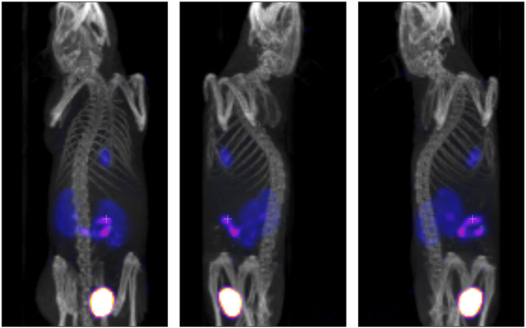
Example MIP projections at three different angles are shown below: